
Dick Vet Equine Hospital first in Scotland to offer standing CT and MRI to patients
The University of Edinburgh’s Dick Vet Equine Hospital in Midlothian Science Zone, has taken delivery of a new standing CT scanner to complement its existing standing MRI scanner for scanning the distal limbs of horses.
The Hospital now offers a further level of imaging modalities for horses referred with lameness and foot problems. The dual imaging modalities will significantly enhance existing diagnostic and treatment expertise at the Hospital.
A three-year study will also assess which imaging systems are preferable for detecting different distal limb problems.
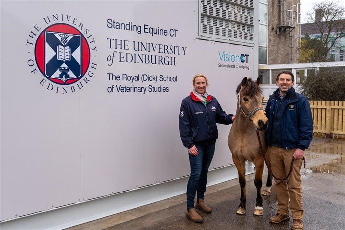
A standing computed tomography (CT) scanner, designed for scanning the distal limbs in the standing sedated horse, was installed at the the Hospital on the University of Edinburgh's Easter Bush Campus in March 2024. The referral hospital already performs CT scans of heads and necks in standing horses and limbs under general anaesthesia. This additional equipment makes the Dick Vet the only equine hospital in Scotland with both a standing MRI and a dedicated standing Equine CT scanner.
Horses with foot and lower limb problems referred to the Hospital for a magnetic resonance imaging (MRI) scan during the three years following installation will receive a CT scan beforehand, at no further cost to the client. The CT scan will be used to check the hoof wall for metal clenches that may be present prior to MRI, to avoid any injury associated with metal migrating in the MRI. The Hospital currently use X-rays for this process. This multi-modal, three-dimensional (3-D) imaging approach using both MRI and CT, will also provide an enhanced level of information about the patient’s hoof capsule and its contents, to aid diagnosis and treatment.
Using advanced imaging technology, the Hospital’s new CT scanner rotates around the patient’s leg to perform the high resolution 3-D scan in as little as three minutes. The cone beam nature of this device means it can be programmed to focus on a specific area of the lower part of the leg, minimising radiation exposure to any surrounding tissues.
The scanner is housed in a purpose-built room. With a walk-in, walk-out floor-level system, easy entry and safe exit is ensured for standing, sedated horses, removing the need for general anaesthesia.
Both CT and MRI are considered advanced imaging modalities and can provide 3-D imaging of the lower limbs. However, each has different strengths when diagnosing foot problems in horses.
An MRI scan is based on the movement of protons. Tissues that have very tightly bound protons, such as bone, normal tendons and ligaments and the hoof wall, have low signal (and appear black) on low field MRI, as the lack of proton movement produces very little signal to generate an image. MRIs are therefore better at showing soft tissue damage to tendons and ligaments when the protons can move in the inflammatory fluid and appear with increased signal (white) as well as active changes in the bone such as bone oedema.
CT relies on X-rays and tissue density, so is better at identifying issues affecting the bone and hoof wall. A standing CT scan can also image the foot, pastern and fetlock simultaneously in under five minutes, once the patient is sedated and position whereas an MRI of these areas would take 2-3 hours.
The most common reason a horse is referred to a vet is lameness, often originating in the foot. The Orthopaedic Service at the Dick Vet Equine Hospital sees a large number of referred horses with lameness and foot problems each year.
With a large team of clinicians, including European and American Diplomates in surgery, medicine, dentistry, anaesthesia, neurology, behaviour, sports medicine, dermatology and ophthalmology, the Dick Vet Equine Hospital is one of the UK’s leading equine referral hospitals. The orthopaedic team work closely with on-site farriers to optimise treatment of foot-related lameness.
As MRI and CT scanners work in different ways, it can be difficult to determine which of these imaging modalities is best to identify different problems of the equine distal limb.
The new scanner, which is a Hallmarq VisionCT, along with the existing Hallmarq MRI, will enhance the technical capabilities of the Hospital, which is one of the most advanced facilities in Scotland.
Specialists at the Hospital will also use the new CT scanner to help work out if MRI or CT or both MRI and CT are necessary for diagnosis of the different conditions of the equine foot. The three-year project will use data from anonymised images of its patients to compare the sensitivity and specificity of the different CT and MRI systems for both navicular syndrome, sometimes known as podotrochleosis, and coffin joint osteoarthritis. The results will help inform vets which system to use to identify the different conditions diagnosed within the equine distal limb. This information will also help minimise ‘over-imaging’ of patients in the future.
“We are excited to offer both standing MRI and CT scans at no extra cost to our clients. This will significantly aid the diagnosis of lameness of our patients. Having both imaging modalities will also provide an excellent opportunity to do some sensitivity and specificity studies to determine whether CT or MRI is better for detecting different injuries in a horse’s foot.”
“Having used the Hallmarq MRI system for over 19 years at the University of Edinburgh I am looking forward to seeing the additional benefits combined CT and MRI imaging can bring to lameness diagnosis.”
For further information, please contact:
The Royal (Dick) School of Veterinary Studies,
The University of Edinburgh, Easter Bush Campus, Midlothian EH25 9RG
Deputy Head of School - Operations:
Cat Eastwood